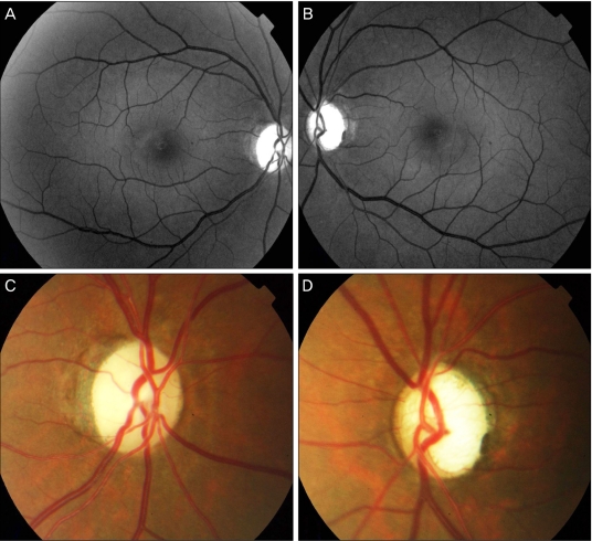
Fig. 2.
The patient presented to our hospital four years after the initial visit for methanol intoxication. (A,B) Red free photographs of both eyes. Red free photographs demonstrate diffuse defects of retinal nerve fiber layers in both eyes. (C) Disc photograph of the right eye, which shows optic nerve atrophy. (D) Disc photograph of the left eye which shows severe disc cupping.

