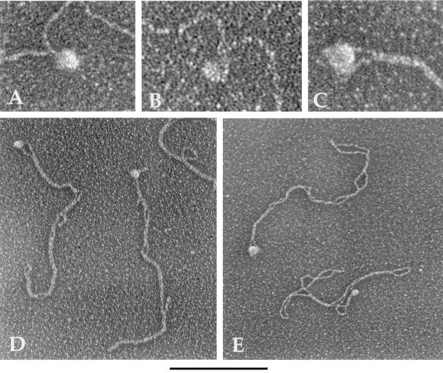
Figure 5.
Visualization of type II topoisomerase molecules bound to relaxed and (−) supercoiled DNA. pBR322 DNA was incubated with yeast topoisomerase II (A, B, D) or E. coli topoisomerase IV (C, E), as described in the text. The DNA was relaxed in A and B and supercoiled in C–E. The complex in C is an enlargement of a molecule in E. After incubation, the samples were prepared for EM by fixation and rotary shadowcasting with tungsten. Images are shown in reverse contrast. [Bar = 0.33 kb of DNA (A–C) and 1.0 kb of DNA (D, E).]

