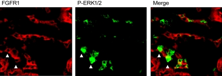
Figure 3.
Localization of p-ERK1/2 and FGFR1. FGFR1 (red) staining was detected in the kidney (left panel). Staining for p-ERK1/2 (green, center panel) colocalized with a subset of FGFR1-positive cells in FGF23-injected mice. Arrows highlight cells that are FGFR1 and p-ERK1/2 positive in the same kidney section.

