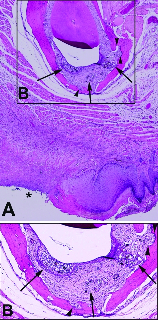
Figure 2.
(A) Cross-section of mandibular incisor hair-induced periodontitis with ipsilateral ulcerative dermatitis (asterisk). Magnification, ×4. (B) Higher magnification of the boxed region of panel A shows expansion of the periodontal space with inflammation, fibrosis, and hair fragments (arrows). Also note the scalloping of mandibular bone due to resorption and reformation (arrowheads). Magnification, ×10.

