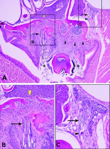
Figure 3.
(A) Cross-section of maxillary molar with extreme, bilateral expansion of the gingival sulcus. Massive amount of hair fragments (asterisk) surrounded by granulomatous inflammation and fibrosis. Also note the scalloping and discontinuity of the maxillary bone (yellow arrowheads) as well as the amount of bone resorption and new formation (arrowheads). Magnification, ×4. (B) Higher magnification of the boxed area labeled B in panel A, showing hair-induced fibrosis and bone resorption and reformation (arrow). Magnification, ×40. (C) Higher magnification of the boxed area labeled C in panel A, showing hair-induced inflammation with multinucleated giant cells (arrowhead). Magnification, ×40.

