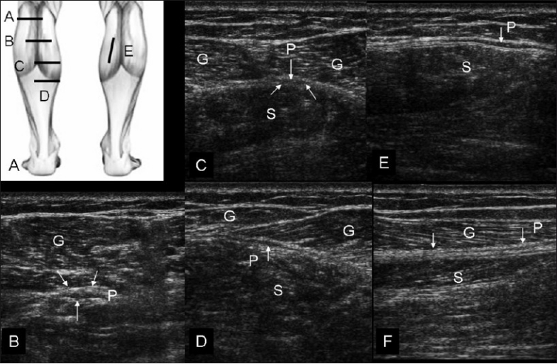
Figure 2 (A–F).

Normal sequential transverse scans of the calf, with the probe positions shown in (A). In position (B), obtained at a relatively high level, the small plantaris (P) muscle is seen below the lateral head of gastrocnemius (G). With progressive distal scans (at levels C, D, and E), the thin tendon of the plantaris (P) (arrows) is seen passing through the triceps surae plane. (F) shows longitudinal view of the plantaris tendon (arrows, P)
