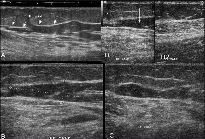
Figure 5 (A–D).

Case 3. Extended-field-of-view longitudinal (A) and standard longitudinal images (B-D1) show a similar hematoma (but with an irregular shape) (arrows); the adjacent muscles show disruption of the normal pennate appearance. Comparison with the normal side (D2) shows that the gastrocnemius has an edematous appearance, with loss of the pennate appearance
See Video 2 at www.ijri.org: Video 2: Case 3. Real-time video gives a better perspective of the findings: hematoma & muscle changes.
