
Figure 11 (A–C).
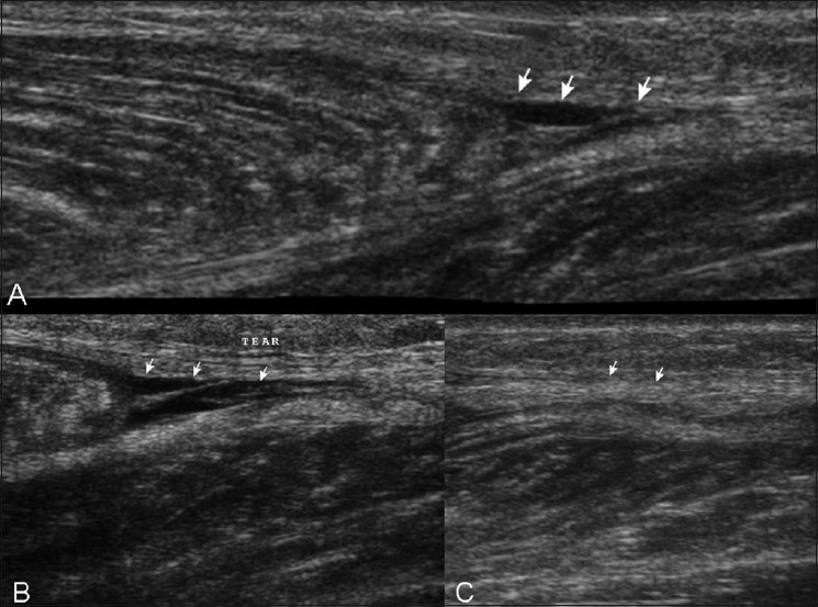
Case 10. Extended-field-of-view longitudinal (A), standard longitudinal (B), and transverse (C) images shows a small hematoma (arrows) near the distal aponeurosis and minimal retraction of the medial head of the gastrocnemius
See Video 3 at www.ijri.org: Case 10. A longitudinal sonogram showing small hematoma near the distal aponeurosis
See Video 4 at www.ijri.org: Case 10. Transverse sonogram in the same case showing infero-superior sweep with similar findings of hematoma near distal aponeurosis