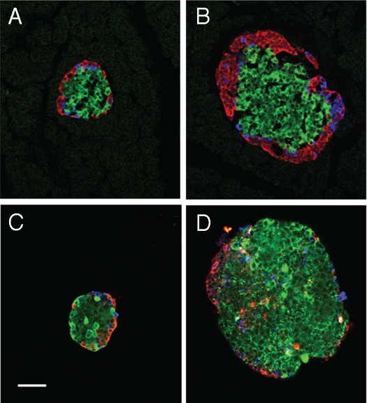
Figure 2.
Cellular composition does not differ with islet population. Islets were immuno-fluorescently labeled for β-cells (insulin = green), α-cells (glucagon = red) and δ-cells (somatostatin = blue). Small (A) and large (B) islets labeled within pancreatic sections (in situ) show same general cell composition. Small (C) and large (D) islets after isolation also have the same general composition. Loss of peripheral α- and δ-cells was noted in the isolated islets (C and D) compared to in situ (A and B). (Scale bar = 50 µm for all images).

