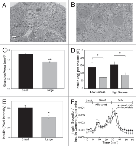
Figure 4.
Small islets have greater insulin content than large islets. (A) Typical TEM micrograph showing β-cells from small islet with densely packed insulin granules. (B) Typical β-cells from large islet with fewer insulin granules. Scale bar = 2 µm for images (A and B). (C) β-cells from isolated small islets have a greater density of insulin granules than β-cells from large islets (p < 0.001). (D) Total insulin content from small (black bars) and large (gray bars). Isolated islets (measured by ELISA) showed that small islets, in low or high glucose, contained more insulin per volume (p < 0.05). (E) Insulin labeling intensity of islets in situ also demonstrated higher values for β-cells from the small islets compared to large islets (p < 0.05). (F) After normalizing insulin secretion to total insulin content/islet, there was no statistical difference in the level or timing of the first or second phase insulin secretion amount between large and small islets.

