Figure 5.
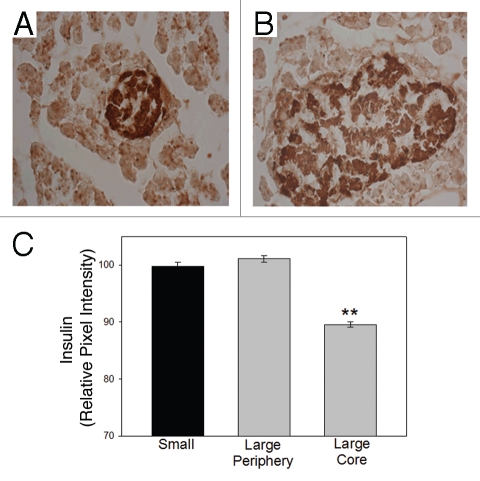
Core β-cells of large islets have less insulin than peripheral β-cells in situ. (A) Typical small islet showing dark insulin staining (brown) throughout the islet. (B) Typical large islet contains lighter insulin-stained β-cells at the core of the islet. (C) Analysis of single cells illustrates that the core β-cells of the large islets contain less insulin/cell. (**p < 0.001).

