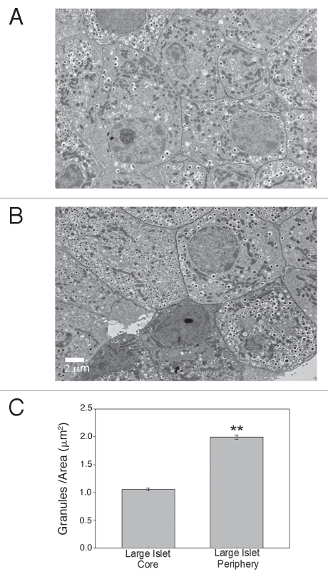
Figure 6.
Core β-cells of large islets have less insulin than peripheral β-cells in vitro. (A) Typical β-cells from core of large islet with few insulin granules/area. (B) Typical β-cells from periphery of large islet with a higher density of insulin granules. (Scale bar = 2 µm for both images) (C) Analysis of cells from core and periphery of large islets showed statistically greater insulin granules/area in the peripheral region. (**p < 0.001).

