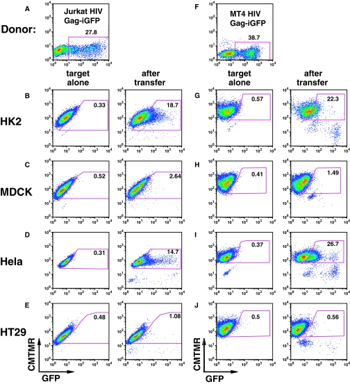
Figure 2.
HIV-1 transfer occurs between HIV-expressing T cells and some epithelial cell lines. (A) HIV Gag-iGFP–transfected Jurkat cells were used as donor cells to epithelial cells below. (B) HK-2 cells were examined before (left) and after (right) 3-hour co-culture with HIV Gag-iGFP–nucleofected Jurkat cells. (C) MDCK cells, (D) HeLa cells, and (E) HT29 cells as target cells with HIV Gag-iGFP–nucleofected Jurkat cells. (F) HIV Gag-iGFP–infected MT4 cells were used as donor cells to the epithelial cells below. (G) HK-2 cells exposed to HIV Gag-iGFP–nucleofected Jurkat cells. (H) MDCK cells, (I) HeLa cells, or (J) HT29 cells as target cells with MT4 cell donors. Epithelial cells were labeled with CMTMR orange and grown in a 24-well plate overnight. Following co-culture for 3 hours at 37°C, cells were washed three times with PBS, detached with trypsin-EDTA treatment at 37°C for 5 to 8 minutes, fixed with 4% PFA, and analyzed by flow cytometry. Numbers within the gate show the percentage of target cells acquiring GFP signal from the donor cells.

