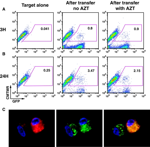
Figure 8.
Exposure of primary tubular cells to primary T cells infected with HIV NL-GI results in GFP expression in epithelial cells at 24 hours. (A) FACS profile of GFP fluorescence in epithelial cells after 3 hours of co-culture. (B) FACS profile after 24 hours of co-culture. (C) Flow sorted GFP-expressing primary renal epithelial cells examined by confocal microscopy show both diffused and punctate dots of GFP in the cytoplasm. DAPI staining of cell nuclei (blue), GFP expression (green), and CMTMR (red).

