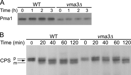
FIGURE 9.
Vacuolar proteolysis in vma3Δ strain. Wild-type and vma3Δ cells were pulse-labeled with Expre35S35S for 5 min and chased for various times. Pma1 (A) and CPS (B) were immunoprecipitated from lysate and analyzed by SDS-PAGE and fluorography. The position of precursor (p) and mature (m) forms of CPS are indicated by black lines. The arrow indicates an intermediate band position where pCPS and mCPS forms comigrate (55).

