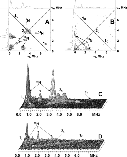
FIGURE 4.
Contour (A and B) and stacked (C and D) presentations of the HYSCORE spectra of the QH SQ with 13C-labeled methyl and methoxy groups in uniformly 15N-labeled D75E and D75H enzymes (magnetic field 346.2 mT (D75E) and 345.7 mT (D75H), time between first and second pulses τ = 136 ns, microwave frequency 9.706 GHz (D75E) and 9.697 GHz (D75H)).

