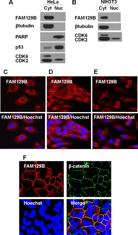
FIGURE 2.
Intracellular localization of FAM129B in exponential and confluent cell cultures. A, HeLa cells (5 × 10⁶) were harvested during the late exponential growth phase, and the cell extracts were fractionated into cytoplasmic (Cyt) and nuclear (Nuc) fractions (as described under “Experimental Procedures”). The fractions were analyzed by immunoblotting using antibodies directed against FAM129B, PARP, p53, CDK6, and CDK2. B, the same procedure was used to analyze NIH3T3 cell fractions. C, immunofluorescence microscopy of exponentially growing HeLa cells stained with a Hoechst 33342 DNA stain and with antibodies directed against rabbit FAM129B and the secondary antibody, chicken anti-rabbit IgG Alexa Fluor 594. The same protocol was followed for the cells in confluent HeLa cells (D) and an exponentially growing HeLa culture showing some mitotic cells (E). F, immunofluorescence co-localization (as described under “Experimental Procedures”) of FAM129B and β-catenin. The confluent cells were co-stained with rabbit antibodies directed against FAM129B and mouse antibodies directed against β-catenin. The secondary antibodies were Alexa Fluor 594-conjugated anti-rabbit IgG and an Alexa Fluor 488-conjugated anti-mouse IgG antibody. The cells were also stained with Hoechst 33342.

