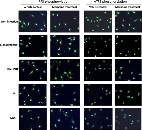
FIGURE 6.
Effect of morphine treatment on phosphorylation of IRF3 and ATF-2 following infection with S. pneumoniae or stimulation with TLR and Nod2 ligands. Immunofluorescence staining reveals the subcellular distribution of IRF3 and ATF2 after infection with S. pneumoniae and stimulation with TLR or Nod 2 ligands. Of note, IRF3 and ATF2 were stained with green labeled secondary antibody. Original magnification was ×400.

