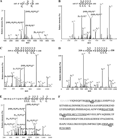
FIGURE 1.
Cdc25B201 LC-MS/MS analysis identified three PKA phosphorylation sites, including Ser149, Ser229, and Ser321. A, MS2 spectrum of the phosphorylated peptide, 147FRS*LPVR153, containing Ser149; S* indicates that serine residue 149 is phosphorylated by PKA. B, MS3 spectrum of the phosphorylated peptide 147FRS#LPVR153, containing Ser149, S# indicates neutral loss of H3PO4 from sequence ions. C, MS2 spectrum of the phosphorylated peptide 319SPS*MPCSVIRPI330 containing Ser321; S* indicates that serine residue 321 is phosphorylated by PKA. D, MS3 spectrum of the phosphorylated peptide 319SPS#MPCSVIRPI330 containing Ser321; S# indicates neutral loss of H3PO4 from sequence ions. E, MS2 spectrum of the phosphorylated peptide 220RQAFTQRPSS*APDLMCLTTEWK241, containing Ser229; S* indicates that serine residue 229 is phosphorylated by PKA. F, amino acid sequence of the Cdc25B201 protein. The LC-MS/MS analysis coverage rate of the given sample is >97% (the covered sequence is shown in boldface letters). The underlined fragments indicate the identified phosphorylated peptide; pS represents phosphorylated serine, including Ser(P)149, Ser(P)229, and Ser(P)321.

