FIGURE 7.
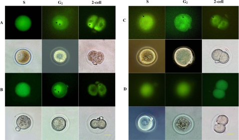
Expression and localization of pEGFP-Cdc25B in the one-cell stage of development of fertilized mouse eggs. A, fertilized mouse eggs injected with pEGFP-Cdc25B-WT. B, fertilized mouse eggs injected with pEGFP-Cdc25B-S149A. In these two groups, the green fluorescent signals were observed in the whole cell at the S stage, and much stronger signals were detected in the nucleus at the G2 phase (double arrow). However, in the two-cell stage, green fluorescent signals were mainly detected in the cytoplasm (arrow). C, fertilized mouse eggs injected with pEGFP-Cdc25B-S149D. The green fluorescent signals were detected in the whole cell but were much more obvious in the cytoplasm (arrow). D, eggs injected with pEGFP-C3. The green fluorescent signals diffused evenly throughout the whole cell, and there was no difference among the various phases of the cell cycle. Scale bar, 20 μm.

