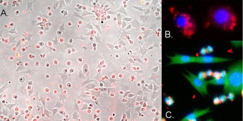
FIGURE 2.
Uptake of CrV2 by specific P. rapae hemocytes. Pieris larvae were bled into media saturated with phenylthiourea; the hemocytes were applied to the wells of a glass slide and allowed to adhere and spread. CrV2 in media was then applied to the cells and incubated for 45 min. A, merged image of bright field and red fluorescence from TRITC secondary antibody bound to anti-CrV2 shows CrV2 associated with the small round hemocytes. B, merged blue and red fluorescence images showing the nucleus (blue) stained with DAPI and CrV2 (red). C, merged blue, red, and green fluorescent image showing the nucleus (blue), CrV2 (red), and the cytoskeleton stained with FITC-conjugated phalloidin (green).

