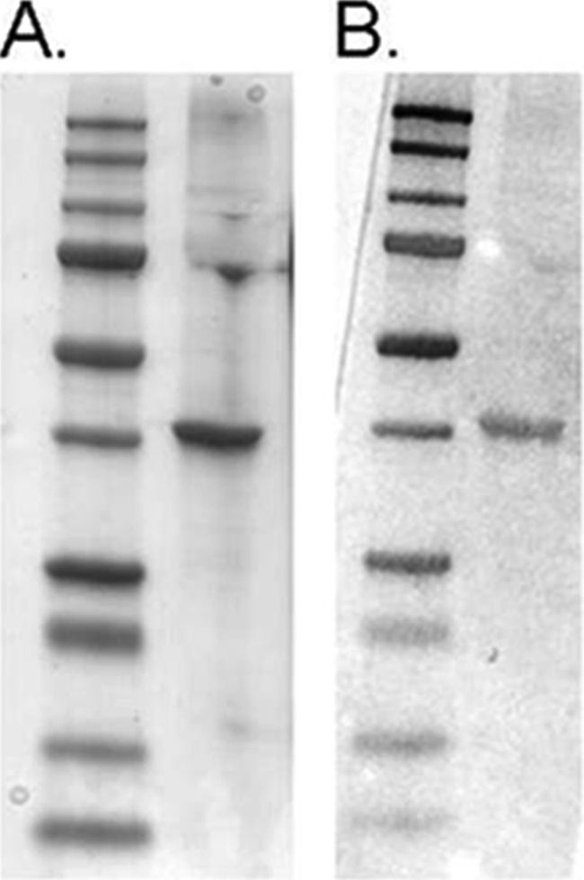
FIGURE 8.

Far Western blot confirms Gαo interaction with CrV2. A, Coomassie-stained gel of Gαo (right lane) run by SDS-PAGE with molecular weight marker (Bio-Rad precision plus dual color protein standard) in left lane. B, duplicate samples of A transferred to nitrocellulose and probed with CrV2. CrV2 binding was detected using rabbit anti-CrV2 and alkaline phosphatase-conjugated secondary antibody. CrV2 bound to the band of Gαo but did not bind to BSA under the same conditions (data not shown). Gαo was not detected when probe CrV2 or anti-CrV2 was absent (data not shown).
