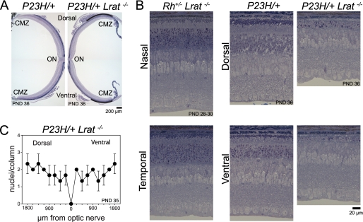
FIGURE 10.
Effect of genetic depletion of 11-cis-retinal production on retinal degeneration. A, whole retinal images obtained from PND 36 P23H/+ and P23H/+Lrat−/− littermate mice. Plastic sections were made from dorsal-ventral axis cut and stained with toluidine blue. Both mice were heterozygous with respect to the L450M variation of RPE65 (RPE65450L/450M). The ONL, observed as a purple line in the P23H/+ mouse retina (left panel), was almost gone and only retained near the cilial marginal zone (CMZ) in the P23H/+Lrat−/− mouse (right panel; indicated by curved line). B, high magnification retinal images shown in A and Rho+/−Lrat−/−mice at PND 28–30. Images were taken from the midpoint between optic nerve (ON) and ciliary marginal zone. Genetic deletion of 11-cis-retinal production promoted retinal degeneration in the P23H/+ mouse at PND 36 but not in Rho+/−/Lrat−/− mouse at PND 28–30. In the P23H/+Lrat−/− mouse, however, the ROS were almost undetectable, and the number of photoreceptor nuclei was dramatically reduced compared with a littermate that had 11-cis-retinal (P23H/+). C, statistical data of the number of photoreceptor nuclei per ONL column in P23H/+Lrat−/−. Cryosections from PND35 P23H/+Lrat−/− RPE65450L/450L− mice were used for quantification. Data were derived from three eyes of three mice. P23H/+Lrat−/− mice showed severe retinal degeneration, and numbers of photoreceptor nuclei per column were similar to those in the P23H/P23H mouse (Fig. 3B, PND 35).

