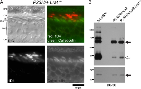
FIGURE 11.
Subcellular localization and protein levels of opsin after genetic deletion of 11-cis-retinal production in Lrat−/− mice. A, cryosections of retina from P23H/+Lrat−/− mice were subjected to immunohistochemical analyses with antibodies against opsin (1D4) and calreticulin at PND 35. Even though massive photoreceptor degeneration is seen, subcellular localization of opsin was normal. B, retinal homogenates from mice at PND 14 were labeled with N-terminal (B6-30) opsin antibodies. All lanes were loaded with 2 μg of homogenate. hrhoG/+ denotes the sample from a heterozygous GFP-tagged human knock-in opsin (hrhoG) and endogenous WT opsin mouse. P23H/hrhoG, heterozygous hrhoG and P23H mouse. P23H/hrhoG Lrat−/−; a heterozygous hrhoG and P23H knock-in mouse lacking lecithin-retinol acyltransferase. GFP-tagged human opsin (∼70 kDa) is indicated by an arrow. Opsin monomers (∼35 kDa) are indicated by an open arrow. Top panel, 4-min exposure was used to analyze the ratio between P23H opsin or WT opsin and GFP-tagged human opsin. Lower panel, 1-s exposure was used to analyze the signal intensity of GFP-tagged human opsin in each lane. The P23H protein level was significantly lower than GFP-tagged human opsin even though 11-cis-retinal production was genetically depleted. DIC, differential interference contrast. RPE, retinal pigmented epithelium; OS, outer segment; IS, inner segment; OLM, outer limiting membrane; ONL, outer nuclear layer; OPL, outer plexiform layer.

