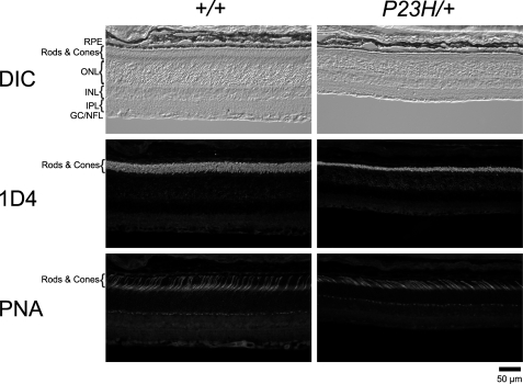
FIGURE 4.
Rods and cones in P23H/+ mice. Cryo-sections from PND 35 WT and P23H/+ mice from the same litter were double-stained with opsin (1D4) antibody and lectin PNA. Images labeled with same marker were captured under identical conditions. Significant shortening of ROS was observed in P23H/+ mice, but the number of cone photoreceptor cells labeled by PNA was not reduced by this age. RPE, retinal pigmented epithelium; ONL, outer nuclear layer; INL, inner nuclear layer; IPL, inner plexiform layer; GC/NFL, ganglion cells/nerve fiber layer; DIC, differential interference contrast.

