FIGURE 3.
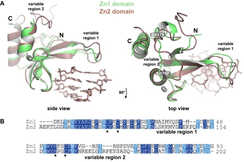
Alignment of the Zn1 and Zn2 domain structures in complex with DNA. A, two views of an alignment of the Zn1 and Zn2 domains, highlighting two structurally distinct regions, labeled variable region 1 (base stacking loop) and variable region 2. Only the DNA duplex from the Zn1-DNA structure is shown for clarity. B, structure-based amino acid sequence alignment of human PARP-1 Zn1 and Zn2 domains. Conserved residues are shaded blue; zinc-coordinating residues are marked with stars.

