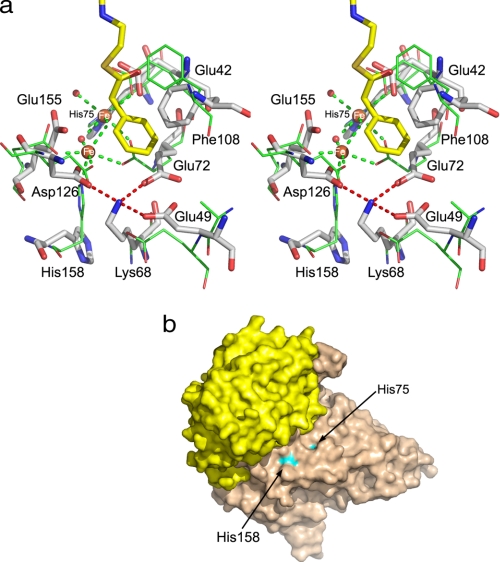
FIGURE 3.
Comparison of substrate-binding sites in MMBs. a, superposition of iron binding centers in PaaA (shown as sticks and labeled) and ToMOH (lines). Glu-72, Glu-155, and His-158 in PaaA in the absence of Fe2+ are rotated away. The “lysine bridge,” Lys-68 salt-bridged to Glu-49, Glu-72, and Asp-126, is also shown. b, surface representation of PaaAC showing that His-75 and His-158 near the di-iron center are partially solvent-exposed.

