FIGURE 1.
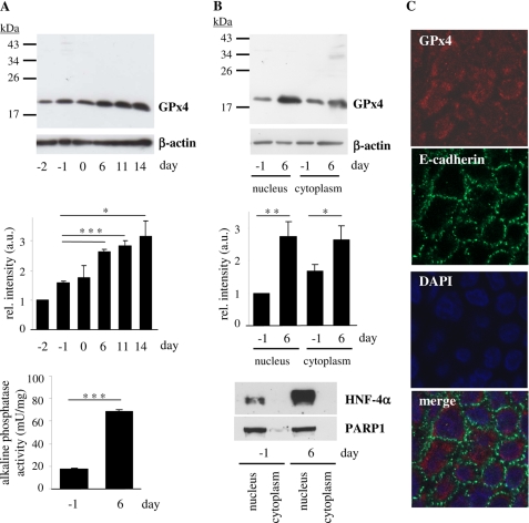
GPx4 expression is stimulated in the nuclear and cytoplasmic compartment of Caco-2 cells in the course of differentiation. Caco-2 cells were cultured for the indicated periods of time with respect to the day reaching confluency (day 0). A, GPx4 protein levels were determined in whole cell lysates by immunoblotting, using β-actin as loading control (upper panel). In the densitometric analysis of GPx4 immunoblots, the data represent means ± S.D. of three independent experiments (middle panel); *, p < 0.05 and ***, p < 0.001. Alkaline phosphatase activity as marker of differentiation was measured in proliferating (day −1) and differentiated (day 6) Caco-2 cells (lower panel). ***, p < 0.001. B, detection of GPx4 protein levels in nuclear and cytoplasmic fractions of proliferating and differentiated Caco-2 cells by immunoblotting (upper panel) and densitometric analysis of three independent experiments (middle panel). *, p < 0.05 and **, p < 0.01. Fractionation was monitored by detection of nuclear proteins HNF-4α and PARP1 (lower panel). C, determination of subcellular localization of GPx4 in differentiated Caco-2 cells (day 6) by immunostaining: GPx4 (red), E-cadherin, a plasma membrane-associated protein (green), nuclei stained with DAPI (blue). mU, milliunits; rel., relative; a.u., arbitrary units.

