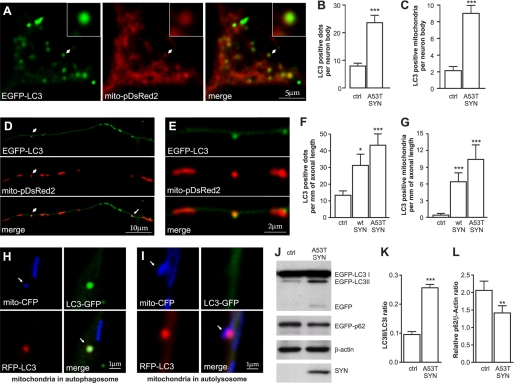
FIGURE 1.
Overexpression of mutant A53T α-synuclein induces mitochondrial removal. Cortical neurons were transfected with plasmids expressing WT or mutant A53T α-synuclein, EGFP-LC3, and mito-pDsRed2 at day 2 after plating and visualized 6 days later. A, colocalization of EGFP-LC3 with mito-pDsRed2 in the cell body of neurons overexpressing mutant A53T α-synuclein. The left panel shows the EGFP-LC3 signal, the middle panel shows the mito-pDsRed2 signal, and the right panel shows the merged signals. The magnified inset shows the colocalization between the mitochondrial and autophagosome signal. Further analysis demonstrated an increased number of autophagosomes (B) as well as an increased number of mitochondria that colocalized with autophagosomes (C) per neuron body in neurons overexpressing mutant A53T α-synuclein overexpressing neurons (Mann-Whitney test). D, upper part of the panel shows the EGFP-LC3 signal, the middle panel shows the mito-pDsRed2 signal, and the lower panel shows the merged signals in neurites of neurons overexpressing mutant A53T α-synuclein. A magnified portion of the picture (E) demonstrates the exact colocalization between the mitochondrial and autophagosome signals. F and G, an increased number of autophagosomes (F) as well as increased number of mitochondria that colocalize with autophagosomes (G) per mm of axonal length in both the WT and mutant A53T α-synuclein overexpressing neurons (Kruskal-Wallis test followed by Dunn's test). H and I, neurons were transfected with A53T α-synuclein, mito-CFP, and RFP-LC3-GFP. H, Mito-CFP and RFP-LC3-GFP colocalize in the autophagosome while in the autolysosome (I) the GFP signal had faded in the acidic environment. J–L, overexpression of A53T α-synuclein enhances LC3-I conversion into LC3-II and decreases the levels of p62 (paired t test). *, p < 0.05; **, p < 0.01; ***, p < 0.001 versus control.

