FIGURE 2.
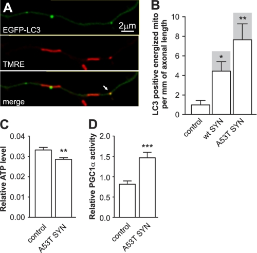
Overexpression of mutant A53T α-synuclein induces a bioenergetic deficit. Cortical neurons were transfected with plasmids expressing WT or mutant A53T α-synuclein and EGFP-LC3 and the mitochondria were stained with the mitochondrial membrane potential sensitive dye TMRE. A, colocalization of EGFP-LC3 (upper), TMRE (middle), and the superimposed channel (lower). B, colocalization analysis demonstrated a considerable increase of LC3-positive energized mitochondria in neurons overexpressing α-synucleins. Also note that the majority of LC3 positive mitochondria were energized (shaded columns show the total number of colocalizations; Kruskal-Wallis test followed by Dunn's test). C, neurons were cotransfected with plasmids expressing firefly luciferase and Renilla luciferase with or without mutant A53T α-synuclein at day 2 after plating. Firefly luciferase activity was measured in living neurons 24 h post-transfection using DMNPE-caged luciferin as a substrate followed by Renilla luciferase measurement (for normalization) in lysed neurons. The expression of mutant A53T α-synuclein decreased the ratio of firefly to Renilla luciferase activities, which indicated a decline in ATP levels. D, neurons were cotransfected with plasmids coding the Gal4-PGC-1α fusion protein, Gal4-UAS-luciferase reporter, and mutant A53T α-synuclein. Increased luciferase activity demonstrates that mutant A53T α-synuclein increases PGC-1α transcriptional activity (t test). *, p < 0.05; **, p < 0.01; ***, p < 0.001 versus control.

