FIGURE 4.
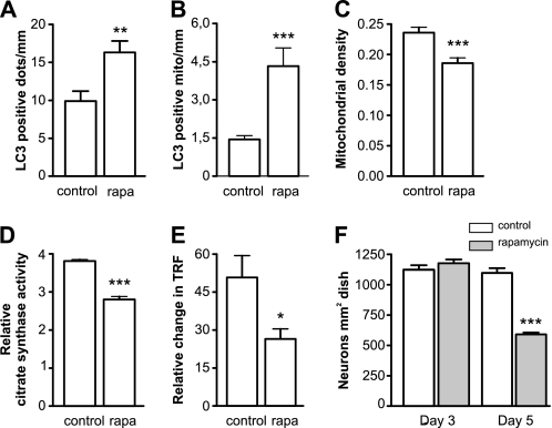
Rapamycin-induced mitochondrial removal and bioenergetic deficit. A and B, neurons were transfected with EGFP-LC3 and mitochondria-targeted pDsRed2 and treated with 100 nm rapamycin at day 2 after plating. Three days post-transfection, the LC3-positive dots were visualized. Rapamycin treatment led to a significant increase in LC3 positive dots (A) as well as LC3 positive mitochondria (B) per mm of axonal length (Mann Whitney test). C, neurons were transfected with GFP and mitochondria-targeted pDsRed2 and treated with rapamycin for 3 days, which led to a decrease in mitochondrial density. Rapamycin-treated neurons had decreased citrate synthase activity (D) and diminished oxygen consumption (E), both of which were normalized per number of intact neurons. Oxygen consumption was estimated by measuring the relative change in time resolved fluorescence (TRF) of the MitoXpressTM probe. F, rapamycin had no effect on neuronal survival after 3 days of treatment, but was clearly toxic after 5 days of treatment. Student's t test, *, p < 0.05; **, p < 0.01; and ***, p < 0.001 versus control.

