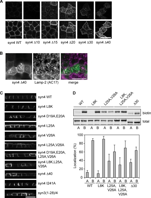
FIGURE 6.
The N-terminal region of syntaxin 4 contains a basolateral sorting signal. A, localization of different deletion mutants in polarized MDCK cells. The different mutants were visualized by staining for GFP. Top images are the apical section and bottom images are the basolateral sections. B, co-localization of syn4-Δ40 mutant with lysosomes. Cells were stained for GFP and Lamp-2, a marker for lysosomes. C, localization of several mutants of syntaxin 4 in polarized MDCK cells. Cells were fixed and stained for GFP. x-z scans of the images are shown. D, localization of syntaxin 4-WT and some syntaxin 4 mutants determined by domain-specific biotinylation at the apical or basolateral membranes. Cells were biotinylated and the biotinylated protein was pulled down using Neutravidin and analyzed by Western blotting with an antibody to syntaxin 4. Top panel shows Western blot. Bottom panel shows the quantification of apical (A) and basolateral (B) localized syntaxin 4 as a percentage of total. The results are shown as the mean ± S.D. of five independent experiments (the difference in localization between syntaxin 4-WT and syntaxin 4-L25A,V26A, -L8K,L25A,V26A, or -Δ30 were all significant with p < 0.01). Scale bars, 20 μm.

