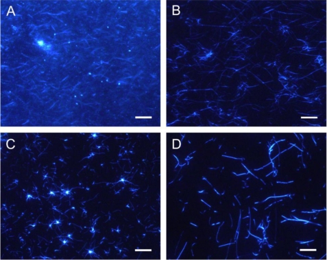
FIGURE 2.
Images of amyloid fibrils of two keratoepithelin peptide fragments observed by TIRFM. TIRFM images of C110–131 (A and C) and H110–131 (B and D) fibrils in the presence of 100 mm NaCl and 0.5 mm SDS. C110–131 (A) and H110–131 (B) fibrils that extended spontaneously in test tubes were observed after incubation for 3 days and 11 days, respectively. C110–131 (C) and H110–131 (D) fibrils that extended seed-dependently on quartz slides were observed after incubation for 6 h. Scale bars = 10 μm.

