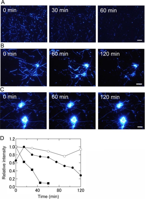
FIGURE 3.
Real-time observation by TIRFM of the destruction of keratoepithelin peptide fibrils by laser irradiation at pH 7. 0 and 37 °C. A, C110–131 fibrils with 2 s of irradiation every 15 min. B, H110–131 fibrils with 2 s of irradiation every 15 min. C, H110–131 fibrils with 2 s of irradiation every 30 min. Fibrils were prepared on quartz slides in the absence of irradiation. Scale bars = 10 μm. D, the time courses of the destruction of these fibrils were obtained by quantifying TIRFM images (see “Experimental Procedures” for details). Shown are C110–131 fibrils with irradiation every 15 min ■, H110–131 fibrils with irradiation every 15 min (●), and H110–131 fibrils with irradiation every 30 min (○). The data points show all of the irradiation applied. The laser power was 80 mW.

