
Figure 5.
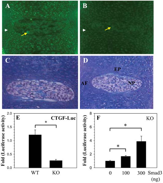
CTGF expression is dependent on Smad3. A-D. Immunohistological expression of CTGF in the intervertebral discs of Smad3 null (KO) and wild type (WT) mice. Saggital sections through the disc of WT mouse embryo (A and C) and Smad3 KO mouse embryo (B and D) were treated with anti-CTGF antibody (C and D) or counterstained with hematoxylin and eosin (C and D). Expression of CTGF was more pronounced in the NP (arrow) and AF (arrow head) of the WT (A) than in the Smad3 KO (B). Mag. × 20. E. CTGF promoter activity in MEF's isolated from smad 3 KO and WT mice. There was an 80% decrease in basal CTGF reporter activity in the Smad3 KO cells F. Smad3 KO Cells were transfected with Smad3 and CTGF reporter activity was measured. A dose dependent increase in CTGF reporter activity was seen with increasing concentration of Smad3. Values shown are mean ± SE from three independent experiments, * p < 0.05.