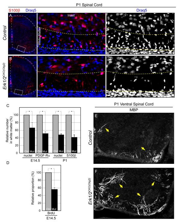
Figure 8. ERK1/2 signaling is necessary for oligodendrocyte precursor proliferation, but is not required for myelination.
A-B. Relative to controls (A) the number of oligodendrocytes in the spinal cord ventral white matter of P1 Erk1/2CKO(Olig2) (B) embryos is strongly reduced.
C-D. Quantification of the relative decrease in the number of nuclei and labeled oligodendrocytes in E14.5 and P1 Erk1/2CKO(Olig2) white matter (C). The proportion of BrdU labeled oligodendrocytes is significantly decreased in Erk1/2CKO(Olig2) E14.5 embryos (D). (mean ± SEM, *=p<0.01)
E-F. At P1, MBP immunolabeling indicates that myelination has just initiated in a small proportion of oligodendrocytes in the ventral spinal cord of control mice (E) (arrows). In Erk1/2CKO(Olig2) (F) littermates, there is a significant increase in the number of oligodendrocytes that express MBP (E) (arrows). (See also Figure S8)

