
Figure 1. Distribution of NCX1 protein in male and female rabbit heart tissue samples were dissected from the epicardium of male and female hearts and processed as described in Methods.
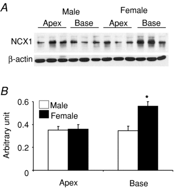
A, Western blot analysis using an NCX1 antibody were compared for protein samples from 3 male and 3 female hearts from the base and the apex of the left ventricular epicardium. The samples were run on the same gel and normalized with respect to their β-actin content. B, summary bar graph of NCX1 protein density from a total of 7 male and 7 female hearts. The data show a statistically significant increase in NCX1 channel protein (P < 0.02) at Mr∼120,000 at the base of female compared to male hearts and the apex of both sexes.