
Figure 7. Effects of E2 on female base endo- and male base epimyocytes.
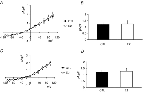
Myocytes were isolated from female base endocardial and male base epicardial regions of rabbit hearts and were incubated with or without E2 (1 nm) for 24 h. A, I–V plots of INCX measured from female base endocardial cells which were incubated in vehicle (DMSO: 1/10,000, n= 5 cells, 3 hearts) or E2 (1 nm, n= 5 cells, 3 hearts). B, INCX (mean ±s.e.m. at 60 mV) was 1.97 ± 0.12 in controls (CTL) and 1.24 ± 0.26 pA pF−1 in oestrogen (E2) treated cells. E2 did not significantly change INCX in female base endocardial myocytes. C, I–V plots of INCX measured from male base epicardial cells which were incubated in vehicle (DMSO: 1/10,000, n= 5 cells, 3 hearts) or E2 (1 nm, n= 5 cells, 3 hearts). D, INCX (mean ±s.e.m. at 60 mV) was 1.20 ± 0.156 in controls (CTL) and 1.25 ± 0.24 pA pF−1 in oestrogen (E2) treated cells. E2 did not significantly change INCX in male base epicardial myocytes.