
Figure 2. Comparison of the longitudinal and lateral transmission of forces in WT and mdx mice.
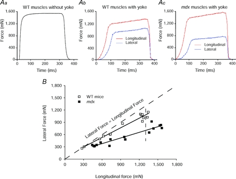
Aa, a record of the maximum isometric tetanic force (Po) of an ATB muscle of a WT mouse. Note ATB and EDL muscles of WT and mdx mice and young and old WT rats display a very similar type of isometric force trace. Ab and c, records obtained following the attachment of the ‘yoke’ apparatus. The forces were measured during maximum stimulation of the motor nerve with the muscle at optimum length for the production of force (Lo). Following the attachment of the ‘yoke’ apparatus, the longitudinal and lateral force traces of maximally activated WT (b) and mdx (c) muscles of mice displayed a slower initial rise in force and then a secondary even slower rise. After the attachment of the ‘yoke’, the Po values of the WT and mdx muscles were ∼25% lower than those of the muscles before the attachment of the ‘yoke’. Note the much closer relationship for the values for the lateral and longitudinal forces for the WT muscle than for the mdx muscle. B, relationship of longitudinal to lateral force transmission in anterior tibialis (ATB) muscles of mdx and WT mice.