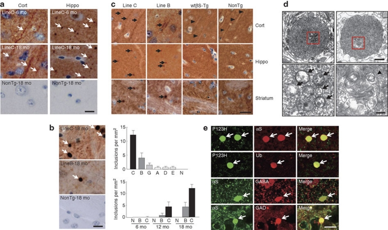
Figure 2. P123H βS tg mice are characterized by a neuritic pathology.
(a, b) Immunohistochemistry of P123H βS using anti-P123H βS antibody. In a, P123H βS accumulated in apical dendrites in the cortex (cort, left panels) and in axonal dots in the hippocampus (hippo, right panels) (6 and 18 months (mo)) of P123H βS tg mice (line C, arrows), but not in the same regions of non-tg littermates (NonTg). Scale bar=10 μm. (b) Globules were observed in the striatum and globus pallidus of 18-month-old P123H βS tg mice (lines B and C, arrows), but not in non-tg littermates. Scale bar=10 μm. For quantification, five images were analysed for each mouse and the results were used to calculate the average number of globules per mm2. Upper panel: multiple lines C, B, G, A, D and E, and non-tg (N) at 18 months; lower panel: lines B, C and N at 6, 12 and 18 months. Data are shown as the mean±s.e.m. (n=7). (c) Immunohistochemistry of total βS using anti-βS antibody. βS accumulated in apical dendrites in the cortex (cort), and in axonal dots in the hippocampus (hippo) of 18-month-old P123H βS mice (lines C and B, arrows), but not in wt βS tg mice (wt βS-Tg) and non-tg littermates (NonTg). Arrowheads indicate negative staining of βS in apical dendrites. Similarly, formation of globules in the striatum was observed in 18-month-old P123H βS mice (arrows), but not in age-matched wt βS tg mice and non-tg mice. Scale bar=20 μm. (d) Photomicrographs of two representative globules in 18-month-old tg mice (line C). The lower two panels are insets from the two higher panels. Globules were composed of small vesicles and contained multivesicular bodies (arrows) and autophagosome-like multiple-layered membranous structures (arrowheads). Scale bar=1 μm (upper panel) and 200 nm (lower panel). (e) Anti-P123H βS-immunopositive globules formed in the basal ganglia of P123H βS tg mice (line C, 18 months) were double stained with either anti-αS or anti-ubiquitin (Ub) (upper six panels). Alternatively, anti-αS-immunopositive globules were stained with anti-αS and either anti-GABA or anti-glutamic acid decarboxylase (anti-GAD) (lower six panels). The slides were observed by laser scanning confocal microscopy. Arrows point to globules. Scale bar=10 μm.

