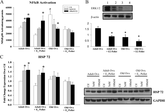
Fig. 3.
NF-κB activation and HSP 72 expression in myocytes from adult and old rats treated with E2 in vitro. A, Graph summarizes the results of NF-κB activation in adult and old rats treated with 5 and 100 nm E2 for 15 min in vitro. B, Graph summarizes ERK 1/2 expression, normalized to β-actin, in the isolated cardiomyocytes. Upper panel, A representative Western blot for ERK 1/2 and β-actin. Lanes: 1, adult ovx; 2, adult ovx + E2 pellet; 3, old ovx; 4, old ovx + E2 pellet. *, P < 0.05 vs. adult ovx. C, Graph summarizes the results of HSP 72 expression in adult and old rats treated with 5 and 100 nm E2 for 12 h in vitro. HSP 72 was normalized to GAPDH expression and to adult ovx. A representative Western blot is shown to the right. n = 5–6 per group and 7–12 plates per group. *, P < 0.05 vs. control (ctl); #, P < 0.05 vs. all other ctls.

