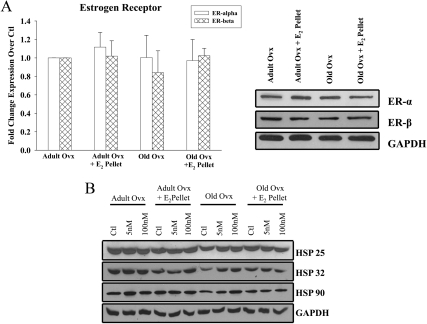
Fig. 4.
ER, HSP 25, 32, and 90 expression in adult and old myocytes. A, Graph summarizes the basal ER-α and β expression in cardiac myocytes from adult and old rats with or without in vivo E2 replacement. ER expression was normalized to GAPDH expression and to adult ovx. Right panel, Representative western blots. n = 3 animals and 3–5 plates per group. Ctl, control. B, Representative Western blots of HSP 25, 32, 90, and GAPDH expression in adult and old rats treated with 5 and 100 nm E2 for 12 h in vitro. n = 6 animals and 6–11 plates per group. P = n.s., data not shown.

