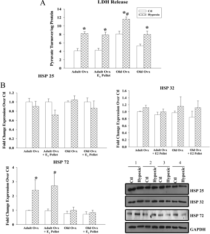
Fig. 6.
Hypoxia-reoxygenation induced release of lactate dehydrogenase (LDH), a marker of cell necrosis. A, LDH release after myocyte hypoxia (12 h) followed by reoxygenation (12 h). B, Graphs summarize HSP 25, 32, and 72 expression in adult and old myocytes with and without E2 replacement under normoxic and hypoxic conditions. All HSP expression was normalized to GAPDH and to adult ovx. Representative Western blots for HSP 25, 32, 72, and GAPDH are shown on the right. Lanes: 1, adult ovx; 2, adult ovx + E2 pellet; 3, old ovx; 4, old ovx + E2 pellet. n = 6 animals and 6–11 plates per group. *, P < 0.05 vs. ctl; #, P < 0.05 vs. all other groups.

