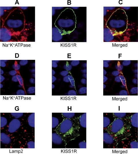
Fig. 2.
Subcellular localization of KISS1R by confocal microscopy. COS-7 cells expressing MYC-KISS1R (Na+K+ATPase colocalization) or CFP-KISS1R (Lamp2 colocalization) were stimulated with 10−7 m kisspeptin for 0–5 h. Anti-CFP antibody was conjugated to FITC (green), whereas membrane (Na+K+ATPase) and lysosome (Lamp2) markers were visualized by Alexa-568-conjugated antibodies (red). Na+K+ATPase (A), KISS1R at baseline (B), and the merged (A + B) image (C); Na+K+ATPase (D), KISS1R after 3 h of kisspeptin stimulation (E), and the merged (D + E) image (F); Lamp2 (G), KISS1R at baseline (H), and the merged (G + H) image (I). Note: D–F, Featured cell is in an upper confocal plan when compared with others. Nuclei are shown in blue (DAPI). Magnification of the images is indicated by the white bar (scale, 20 μm).

