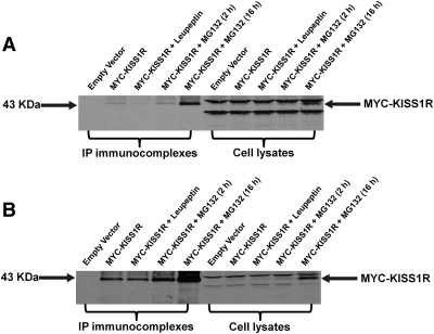
Fig. 4.
Western-Blot detection of MYC-KISS1R. COS-7 (A) or HEK-293 (B) cells expressing MYC-KISS1R were incubated at 37 C with or without leupeptin (lysosome inhibitor) for 16 h or with MG132 (proteasome inhibitor) for 2 h or 16 h before lysis. Cell lysates underwent immunoprecipitation (IP) with a monoclonal anti-myc antibody followed by Western blot analysis. MYC-KISS1R in whole cell lysates or anti-myc immunoprecipitants was detected using a polyclonal anti-myc antibody (1:1,000) followed by a fluorescent-labeled infrared dye IRDye-800CW-conjugated antirabbit antibody (1:5,000). Lanes 1 through 5 were loaded with immunoprecipitated complexes, and lanes 6 through 10 with the corresponding pre-IP whole cell lysates. Lanes 1 and 6, control vector; lanes 2 and 7, MYC-KISS1R alone; Lanes 3 and 8, MYC-KISS1R + Leupeptin (16 h); lanes 4 and 9, MYC-KISS1R + MG132 (2 h); lanes 5 and 10, MYC-KISS1R + MG132 (16 h). Arrow on the right points to KISS1R monomers at 43 kDa. Western blot of representative experiments are shown. Experiments were repeated at least three times with comparable results.

