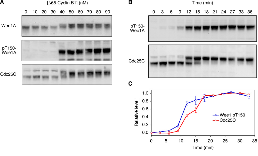
Figure 2. Comparison of Cdc25C and Wee1A Responses.
(A) Steady-state responses. Extracts were incubated for two hours with different concentrations of non-degradable Δ65-cyclin B1, which was sufficient to allow the Wee1A and Cdc25C phosphorylation to approach steady state.
(B, C) Time courses. Extracts were incubated with 100 nM Δ65-cyclin B1 for the lengths of time indicated. Panel B shows one experiment; panel C shows average results from two independent experiments.

