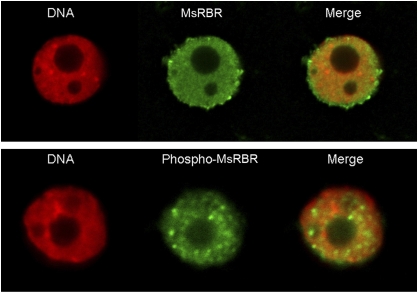
Fig. 7.
Differences in immunofluorescence labelling of MsRBR and phospho-MsRBR proteins in isolated nuclei released from alfalfa cells indicate different nuclear organization for the two forms of MsRBR protein. Upper panel: diffused, uniform staining of nucleoplasm (green) after immunolocalization with anti-AtRBR1 antibody. Lower panel: granular labelling of the nucleus (green) after immunolocalization with anti-human pRb phosphopeptide antibody. DNA stained with DAPI and pseudocoloured red.

