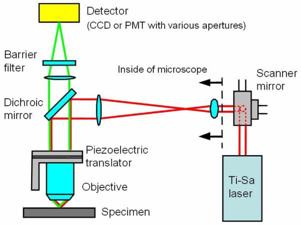
Fig. 1.
An experimental setup for quantifying photon scattering effects on PSFem. Excitation beam path is very similar to conventional multiphoton microscopy. For measuring the PSFem FWHM variation, a CCD camera is used as a detector. For measuring the PSFem spatial distribution, a pinhole aperture of various sizes is positioned in the image plane and a PMT behind the aperture collects the transmitted emission light.

