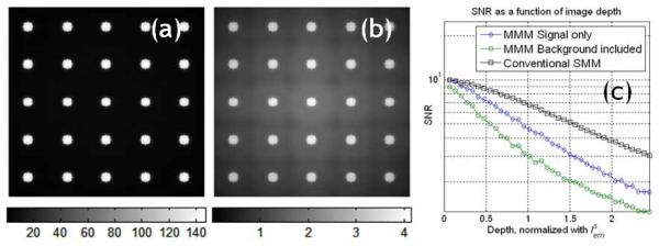
Fig. 4.
A simulation of photon scattering effect on image contrast in MMM tissue imaging. An original image comprising 5 × 5 microspheres of 10 μm diameter equally spaced with 40 μm separation distance and the experimentally measured PSFtot was used in the simulation. (a) MMM image at the sample surface, (b) MMM image at imaging depth of , where increased background noise is observed in the center of the image. (c) The decay of SNR of the MMM image at its center location as a function of the imaging depth.

