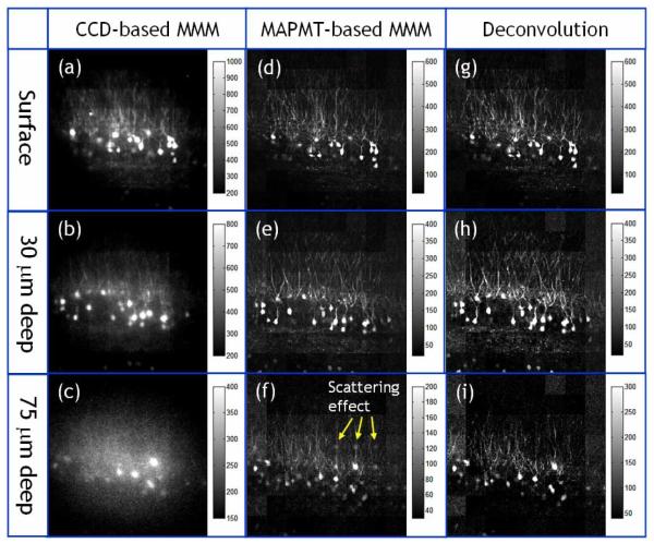
Fig. 9.
Images of GFP expressing neurons in the ex vivo mouse brain acquired with CCD-based MMM (a-c) and MAPMT-based MMM (d-f) at different depth locations (surface, 30 um, and 75 um deep). For images with MAPMT-based MMM, a deconvolution algorithm was applied to remove the effect of emission photon scattering (g-i). The objectiveused is a 20× water immersion with NA 0.95. The input laser power is 300 mW at 890 nm wavelength. The frame rate is 0.3 frames per second with a frame size of 320 × 320 pixels.

